Adam and Eve? Or Alanine and Serine?

What if the defining reason for life on Earth is the manipulation of time? While organic matter has been discovered in outer space, in the form of methane in the ethers of Neptune and Uranus, and asteroids and meteorites that crashed into Earth (Organic Compounds in the Solar System, 2016), why have we not yet discovered extraterrestrial life in the form of a cell that is moving, eating, growing, changing, and replicating (Sagan & Sagan, 2018)?

The defining feature of life could be the enzyme: proteins made from amino acids that increase the rate of chemical reactions. Metabolic reactions could not occur without enzymes (Lewis & Stone, 2020), as reactions would occur too slowly to interact with one another. Enzymes increase the speed of reactions so that complex, multifaceted interactions can occur to create and sustain life. It is the enzyme that has catalyzed life on Earth, by shortening the time needed for reactions to take place.

Although amino acids have been discovered in some meteorites, such as the simpler alanine and glycine found in class CI and CN meteorites (Organic Compounds in the Solar System, 2016), the 20 main ones that define life on Earth have not yet been found elsewhere in the universe in same concentrations and varieties.

There is much debate surrounding the origin of the 20 amino acids. Is it by chance or by evolutionary design? And are more amino acids evolving or could they evolve? Other amino acids do exist, including selenocysteine which is found in humans, but is comparatively more complex to utilize, which might be a clue as to why there aren’t more than the 20 main ones that exist (Brazil, 2017).

Francis Crick hypothesized in his frozen accident theory that 20 specific amino acids exist by chance and that any other number of structures could have been used to synthesize proteins (Koonin, 2017). However, in 2017 Doig makes strong arguments that the 20 main amino acids with their specific hydrophobicity enable folding, stability, accessibility of active sites, and metabolic efficiency, making amino acids the perfect building blocks for proteins and enzymes (Doig, 2017).

Why are C, H, N, O, and S the organic molecules that compose amino acids? In addition to being bountiful on our planet, these elements are ideal for many reasons. In contrast, metals such as selenium and antimony are too soluble in water and therefore unstable. Halogens are too electronegative and reactive. Silicon bonds too readily to oxygen in place of other elements. The list goes on and on to account for the specificity that explains why our current amino acids are the perfect constituents of life (Doig, 2017).

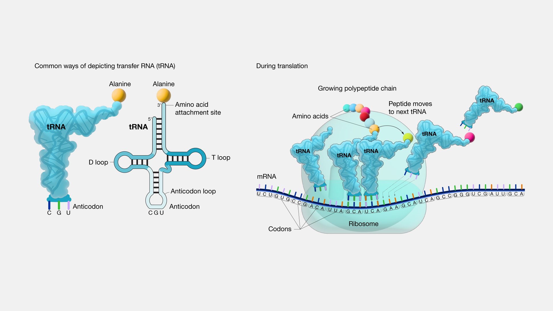
So, if it is not a frozen accident then why has the number of amino acids stopped at approximately 20 when so much of evolution is characterized by seemingly endless diversity? One fascinating theory explains it in terms of simple logistics. tRNA (transfer RNA) reads the genetic code from RNA, and then using the enzyme aminoacyl tRNA synthetase, selects which amino acid tRNA should bind to. tRNA then transfers the selected amino acid to a ribosome for assembly on the growing polypeptide chain. tRNA has only 3 reading sites, and after accounting for the start and stop codons, 61 possible amino acids reading codes. However, tRNAs are limited in their recognition ability (Saint-Léger et al., 2016). tRNAs already make on average one mistake for every 1000-10,000 codon readings and adding more amino acids would enable more mistakes. Dr. Ribas explains it this way, “It’s like if you have a very simple kind of lock where you could only change three or four pins, you come to a point where you wouldn’t be able to make new keys because a new key will open a lock you have already used and that defeats the purpose” (Brazil, 2017).

Photo Credit: (National Human Genome Research Institute, 2019)

Scientists do not have all the answers on whether amino acids evolved by chance or limited necessity, but one thing is certain; the 20 amino acids are brilliant in their specificity and enable life as we know it. Without proteins, we would not have enzymes, and without enzymes, we would still be a pool of RNA and co-factors. Enzymes alter the speed of reactions and amino acids are the specific building blocks of all proteins and enzymes. Without our 20 amino acids, we would lack the complex web of reactions needed to create life; organisms that can move, eat, breathe and reproduce.


REFERENCES

Organic Compounds in The Solar System. (2016, May 26). Chemistry LibreTexts. https://chem.libretexts.org/Ancillary_Materials/Exemplars_and_Case_Studies/Exemplars/Physics_and_Astronomy/Organic_Compounds_in_The_Solar_SystemLinks to an external site.

Sagan, D., & Sagan, C. (2018). life | Definition. In Encyclopædia Britannica. Retrieved from https://www.britannica.com/science/lifeLinks to an external site.

Lewis, T., & Stone, W. L. (2020). Biochemistry, Proteins Enzymes. PubMed; StatPearls Publishing. https://www.ncbi.nlm.nih.gov/books/NBK554481/#:~:text=Enzymes%20are%20proteins%20comprised%20ofLinks to an external site.

Brazil, R. (2017). Why are there 20 amino acids? Chemistry World. https://www.chemistryworld.com/features/why-are-there-20-amino-acids/3009378.articleLinks to an external site.

Doig, A. J. (2017). Frozen, but no accident – why the 20 standard amino acids were selected. The FEBS Journal, 284(9), 1296–1305. https://doi.org/10.1111/febs.13982 Links to an external site. 

Koonin, E. (2017). Frozen Accident Pushing 50: Stereochemistry, Expansion, and Chance in the Evolution of the Genetic Code. Life, 7(2), 22. https://doi.org/10.3390/life7020022 Links to an external site. 

Saint-Léger, A., Bello, C., Dans, P. D., Torres, A. G., Novoa, E. M., Camacho, N., Orozco, M., Kondrashov, F. A., & Ribas de Pouplana, L. (2016). Saturation of recognition elements blocks evolution of new tRNA identities. Science Advances, 2(4). https://doi.org/10.1126/sciadv.1501860Links to an external site.

National Human Genome Research Institute. (2019). Transfer RNA (tRNA). Genome.gov. https://www.genome.gov/genetics-glossary/Transfer-RNA

The past and future of aminoacyl-tRNA synthetases

I chose aminoacyl-tRNA synthetases (aaRSs) to study today because these enzymes seemed like some of the more complex and mysterious molecules from this week’s lectures on protein synthesis. The more I researched, the more I understood how one could completely dedicate one’s life to studying only one enzyme. I read an in-depth paper by Rubio Gomez and Ibba (CSHL Press) and was surprised to discover that there are currently 23 known aaRSs. In addition to the 20 that Goodsell references that each code for a specific amino acid, there are two that code for lysine and two called pyrrolysyl-tRNA synthetase and phosphoseryl-tRNA synthetase found in some archaea and bacteria (Goodsell) (Rubio Gomez and Ibba).

Interestingly, aaRSs presents some interesting questions regarding their evolution, and, as others have mentioned, there are rare genetic disorders linked to aaRs mutations, as well as recent aaRs-targeted drug developments. Interestingly, these enzymes play a central role in the latest biosynthetic research as engineered amino acids are being written into novel polypeptide chains, altering the genetic code and resulting in the creation of biosensors, biomarkers, innovative functioning proteins, viral defenses, and more. Scientists have altered some aaRs:tRNA pairing to associate a new amino acid into translation (Rubio Gomez and Ibba) (Rovner et al.). As there is so much to discuss regarding aminoacyl-tRNA synthetases, I will focus on how they evolved and how they are altering evolution.

AaRSs are antiquated, having been inherited from the Last Universal Common Ancestor (LUCA) (Fournier et al.). And, they seem to have been just as complex in LUCA as they are in contemporary organisms (Rubio Gomez and Ibba). Since aaRSs are the readers of the genetic code, but also the genetic code is required to synthesize them, this presents a ‘chicken or the egg’ dilemma (Rubio Gomez and Ibba). As discussed in lecture 39, there are two types of aaRSs, each approaching the tRNA from a different side with their active site, with type I adding an amino acid to the last 2’ tRNA hydroxyl group and type II adding an amino acid to the 3’ hydroxyl on the final tRNA base (Barbaro). Both types also have a difference in their substrate binding methodology. Despite their difference, there is an accepted theory called the Rodin and Ohno hypothesis that both classes arose from the same gene simultaneously from opposing sides. The codons that code for residues for class I active sites are palindromes for class II sites. In other words, codons for class I are anticodons for class II. (Martinez-Rodriguez et al.). And so, two aaRSs arose due to bidirectional reading of mRNA and attached two different amino acids to tRNAs. This created the first protein comprised of more than one amino acid. Later genetic mutation and editing enabled the diversity of present-day aaRSs (Rubio Gomez and Ibba).

Noncanonical amino acids (ncAAs) are artificially synthesized amino acids that alter the genetic code and create genomically recoded organisms (GROs). GROs are created when scientists reassign a codon to a different and sometimes artificially synthesized amino acid (Lajoie et al.). Thus, new aaRSs are required that can recognize ncAAs and attach them to tRNAs that will transport them to ribosomes for protein chain synthesis. For example, Rovner et al. conducted research on an organism that lacked the TAG codon and reassigned TAG to code for a Methanocaldococcus jannaschii tRNA:aminoacyl-tRNA synthetase pairing. The protein was fabricated and stable.

I was curious to understand how aaRSs are altered to bind nCAAs. I came upon a study that described exactly its methodology and provided a helpful infographic. If you take a look at part c 2. (top right), you can see that a library of aaRS variants is generated after the introduction of the novel amino acid. Through mutation, an aaRS will mutate to associate the codon with the ncAA. This will be selected and amplified using PCR technology (Vargas-Rodriguez et al.). I was surprised that ncAAs are created by selective breeding! 

Image: www.ncbi.nlm.nih.gov/pmc/articles/PMC6214156.

I have only scratched the surface of my understanding of aaRSs. From ancient origins to the future of genetic engineering, it seems there is ad infinitum to discover in the study of aminoacyl-tRNA synthetases. 


REFERENCES

Rubio Gomez, Miguel Angel, and Michael Ibba. “Aminoacyl-tRNA synthetases.” RNA (New York, N.Y.) vol. 26,8 (2020): 910-936. doi:10.1261/rna.071720.119

Fournier, Gregory P., et al. "Molecular Evolution of Aminoacyl tRNA Synthetase Proteins in the Early History of Life." Origins of Life and Evolution of Biospheres, vol. 41, no. 6, 2011, pp. 621-632.

Barbaro B, Biochemistry at U of California San Diego. Course Number: Chapter 39 - the Genetic Code” [accessed 2023 Dec 15]

Martinez-Rodriguez, Luis, et al. “Functional Class I and II Amino Acid-activating Enzymes Can Be Coded by Opposite Strands of the Same Gene.” PubMed Central (PMC), 18 June 2015, www.ncbi.nlm.nih.gov/pmc/articles/PMC4528134 Links to an external site..

Rovner, Alexis J., et al. “Recoded Organisms Engineered to Depend on Synthetic Amino Acids.” PubMed Central (PMC), 21 Jan. 2015, www.ncbi.nlm.nih.gov/pmc/articles/PMC4590768 Links to an external site..

Goodsell, David. “PDB101: Molecule of the Month: Aminoacyl-tRNA Synthetases.” RCSB: PDB-101, pdb101.rcsb.org/motm/16. Accessed 16 Dec. 2022.

“Genetically Modified Organisms (GMOs) | Learn Science at Scitable.” Genetically Modified Organisms (GMOs) | Learn Science at Scitable, www.nature.com/scitable/topicpage/genetically-modified-organisms-gmos-transgenic-crops-and-732. Accessed 16 Dec. 2022.

Lajoie, Marc J., et al. “Genomically Recoded Organisms Expand Biological Functions.” PubMed Central (PMC), www.ncbi.nlm.nih.gov/pmc/articles/PMC4924538. Accessed 16 Dec. 2022.

Vargas-Rodriguez, Oscar, et al. “Upgrading aminoacyl-tRNA Synthetases for Genetic Code Expansion.” PubMed Central (PMC), 27 July 2018, www.ncbi.nlm.nih.gov/pmc/articles/PMC6214156.


Beta-galactosidase as indicator of senescent and cancer cells

Yesterday I was listening to a news segment on CBC Links to an external site.about cervical cancer screening results that currently have 6-month delays in parts of Canada due to post-pandemic backup and staff shortages. Coincidently, while researching beta-galactosidase, I discovered that this enzyme is an important biomarker for certain cancers, including ovarian cancer (Fan et al., 2021) and, most recently, has been found to be over-expressed in gastric cancer as well (Kubo et al., 2021). 

Human beta-galactosidase is highly expressed in senescent cells, which are cells that stop dividing but continue to exist and metabolize. Accumulation of senescent cells is an indicator of aging in general, and the presence of these cells is associated with a risk for many diseases including cancer, osteoarthritis, atherosclerosis (arterial plaque build-up), Alzheimer’s, diabetes, and more (Childs et al., 2017) (Chen et al., 2022). Interestingly, cellular senescence seems to be one of the body’s defenses against tumor growth, in that it’s a way for the body to stop the division of cells that have shortened telomeres and may have mutations (Campisi, 2001). Yet, secretions by senescent cells also cause disease (Childs et al., 2017) and even a small number of senescent cell secretions such as cytokines can have a significant impact on cells throughout the entire body and cause or increase disease (Burton, 2009).  And so, the presence of an accumulation of human beta-galactosidase in tissues indicates aging and the onset or development of disease.  

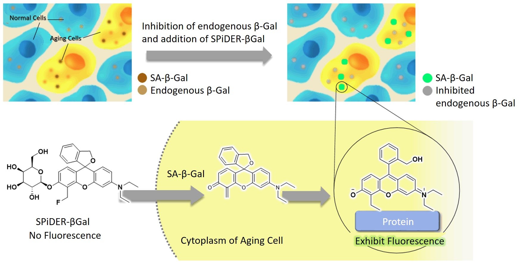
Early cancer detection greatly affects survival rates. Moreover, distinguishing the exact type of cancer aids in treatment decisions. Finding biomarkers that indicate early cancer growth and specify the type of cancer is key to increasing survivorship. Beta-galactosidase is shown to be present in ovarian cancer and most recently associated with gastric cancer (Kubo et al., 2021). Detection of beta-galactosidase has been historically done by a technology called X-gal but has certain disadvantages including that it can only be done in cells that are non-living and the staining takes a long time and is difficult to visualize. However, a new technology called Cellular Senescence Detection Kit - SPiDER-βGal is more sensitive, easier to see, and can be done on live cells (in vivo) (Cellular Senescence Detection Kit - SPiDER-βGal, n.d.).

The image shows beta-galactosidase cells illuminated with the application of SPiDER-βGal technology. Source: (Cellular Senescence Detection Kit - SPiDER-βGal, n.d.).

Research has shown that beta-galactosidase fluorescent illumination is a reliable indicator for gastric cancer, liver, and other cancers. B-galactosidase is found in higher concentrations in tumor tissues compared to healthy cells. The presence of beta-galactosidase can be seen visually, with a stronger fluorescence indicating higher concentrations.  Furthermore, the detection is fast and can be seen only 10 minutes after application. This technology could thus be used during surgery, to help surgeons see cancer cells during surgery, namely fluorescence-guided surgery (Ogawa et al., 2021). Also, it can shine for hours, is non-toxic, and will illuminate even at low concentrations. Detecting and removing even small amounts of cancer cells can greatly decrease reoccurrences or metastasis. 

I became curious to learn exactly how the probe lights up beta-galactosidase and it took hours of research to find some semblance of an explanation. I finally came upon J. Zhang et al., the researchers who originally developed the technology and published their findings, in 2017. The probe is a substrate for Beta-galactosidase and ‘turns on’ in a solution with H2O. It can detect even a small concentration of 0.1 nM and has a very high bonding affinity. The probe can distinguish between endogenous Beta-galactosidase cells and non-endogenous. It has a hemicyanine structure (meaning dyes with two aromatic groups) with a D-galactose residue using a glycosidic bond and s fluorescent intensity at 703nm (J. Zhang et al., 2017). 

I will admit I am still not satisfied with this information and want to really understand how this probe lights up the specific substrate groups. Here is an image that I partially understand. I can see that Gal-Pro is the probe. It meets with Beta-galactosidase. The top sugar (chair-shaped) comes off and binds to beta-galactosidase. I have many remaining questions, such as why it bonds and how it illuminates. I am sure I am limited in my understanding as my organic chemistry knowledge is not comprehensive. Perhaps someone can comment and explain what happens on a substrate level and what causes the fluorescence. 

Image from Qiu et al., 2020.

In summary, beta-galactosidase is present in high concentrations in senescent cells and is an indicator of ovarian and gastric cancer. SPiDER-βGal probe is a powerful new technology that can be used for fluorescence-guided surgery to find and remove cancer cells. It can also be used as a fast biomarker in vivo for the presence of gastric and ovarian cancer, even in small concentrations.


REFERENCES


Kubo, H., Murayama, Y., Ogawa, S., Matsumoto, T., Yubakami, M., Ohashi, T., Kubota, T., Okamoto, K., Kamiya, M., Urano, Y., & Otsuji, E. (2021). β-Galactosidase is a target enzyme for detecting peritoneal metastasis of gastric cancer. Scientific Reports11(1), 10664. https://doi.org/10.1038/s41598-021-88982-2Links to an external site.

Fan, F., Zhang, L., Zhou, X., Mu, F., & Shi, G. (2021). A sensitive fluorescent probe for β-galactosidase activity detection and application in ovarian tumor imaging. Journal of Materials Chemistry. B, Materials for Biology and Medicine9(1), 170–175. https://doi.org/10.1039/d0tb02269aLinks to an external site.

Childs, B. G., Gluscevic, M., Baker, D. J., Laberge, R.-M., Marquess, D., Dananberg, J., & van Deursen, J. M. (2017). Senescent cells: an merging target for diseases of ageing. Nature Reviews. Drug Discovery16(10), 718–735. https://doi.org/10.1038/nrd.2017.116Links to an external site.

Chen, Y.-H., Zhang, X., Ko, K.-Y., Hsueh, M.-F., & Kraus, V. B. (2022). CBX4 regulates replicative senescence of WI-38 fibroblasts. Oxidative Medicine and Cellular Longevity2022, 5503575. https://doi.org/10.1155/2022/5503575

Childs, B. G., Gluscevic, M., Baker, D. J., Laberge, R.-M., Marquess, D., Dananberg, J., & van Deursen, J. M. (2017). Senescent cells: an emerging target for diseases of ageing. Nature Reviews. Drug Discovery16(10), 718–735. https://doi.org/10.1038/nrd.2017.116Links to an external site.

Campisi, J. (2001). Cellular senescence as a tumor-suppressor mechanism. Trends in Cell Biology11(11), S27-31. https://doi.org/10.1016/s0962-8924(01)02151-1

Burton, D. G. A. (2009). Cellular senescence, ageing and disease. Age (Dordrecht, Netherlands)31(1), 1–9. https://doi.org/10.1007/s11357-008-9075-y

Cellular Senescence Detection Kit - SPiDER-βGal. (n.d.). Dojindo.eu.com. Retrieved December 8, 2022, from https://www.dojindo.eu.com/store/p/895-Cellular-Senescence-Detection-Kit-SPiDER-Gal.aspx

Ogawa, S., Kubo, H., Murayama, Y., Kubota, T., Yubakami, M., Matsumoto, T., Yamamoto, Y., Morimura, R., Ikoma, H., Okamoto, K., Kamiya, M., Urano, Y., & Otsuji, E. (2021). Rapid fluorescence imaging of human hepatocellular carcinoma using the β-galactosidase-activatable fluorescence probe SPiDER-βGal. Scientific Reports11(1), 17946. https://doi.org/10.1038/s41598-021-97073-1

Zhang, J., Li, C., Dutta, C., Fang, M., Zhang, S., Tiwari, A., Werner, T., Luo, F.-T., & Liu, H. (2017). A novel near-infrared fluorescent probe for sensitive detection of β-galactosidase in living cells. Analytica Chimica Acta968, 97–104. https://doi.org/10.1016/j.aca.2017.02.039

Qiu, W., Li, X., Shi, D., Li, X., Gao, Y., Li, J., Mao, F., Guo, Y., & Li, J. (2020). A rapid-response near-infrared fluorescent probe with a large Stokes shift for senescence-associated β-galactosidase activity detection and imaging of senescent cells. Dyes and Pigments: An International Journal182(108657), 108657. https://doi.org/10.1016/j.dyepig.2020.108657

What are epigenetics and is it a good or bad thing?

This is in response to this question:

What role does DNA mismatch repair have in cancers? According to the slides this week, "Base-excision repair corrects the most common point mutation in humans, the deamination of methylcytosine to thymine." If this repair does not work, is there an abundant amount of genetic information and does that lead to cancer?

Your question really opens Pandora’s box. The deamination of cytosine into uracil or thymine is a hot topic in epigenetic research. Epigenetics is the theory that environment and experiences change the expression of genes, altering phenotypic expression in somatic and even germ cells. I found a helpful Harvard infographic that quickly explains epigenetics, which I thought might be helpful.

(What Is Epigenetics? The Answer to the Nature Vs. Nurture Debate, n.d.)  

As you can see from the infographic, the original concept of DNA was that it was a blueprint that dictates phenotypic expressions. However, scientists now perceive that there are many regulators and modifiers of DNA that contribute to a diversity of gene expression. Additionally, these modifiers can be passed on from generation to generation.

Studies with rats have shown transgenerational modifications of gene expression based on early maternal-infant relations. For example, increased DNA methylation in the prefrontal cortex that causes depressive behavior is observed in rats abused by their mothers. This methylation is then passed on in subsequent generations and observed in male sperm and female/male somatic cells, along with observed depressive behaviors (Gudsnuk & Champagne, 2012). This methylation was not present in the DNA but is now present as an epigenetic marker influencing gene expression and is then passed on in germ cells. In other words, if a mother rat abuses her baby, it causes methylation in the cortex that causes depression in her baby and her baby’s babies. Epigenetics has a particular influence on the brain due to the plasticity of cerebral neurons.

And so, cytosine methylation into thymine (or uracil) is one of the key players in epigenetics. The transition mutation from cytosine to thymine has many implications when not corrected by base-excision repair, from increased variation in immunological defense to oncogenesis.

In terms of immunity, it is crucial to have antibodies that can recognize and defend against a large and ever-evolving diversity of pathogens. Deamination of cytosine is important for the hypermutation of somatic B cells that produce the necessary antibody diversity. Enzymes such as cytosine deaminases are crucial for an efficient immune system and shown to be lacking in humans with hyper-IgM immunodeficiency. Thus, the deamination of cytosine enables B cells to create the immunoglobin diversity necessary for efficient immunity.  (Chahwan et al. 2012).

Counter to that, deamination mutations of cytosine are also implicated in many cancers. Studies have found that activation-induced cytidine deaminases (AID) cause early stop codons in tumor-suppressor genes in colorectal cancer (Morisawa et al. 2012). Further studies have shown that AID enzymes are present in overabundance in many organ cancers. Although different gene mutations are observed in different cancers, for example, the k-ras gene mutation in pancreatic cancers, and the c-myc gene in lung and lymphoma cancers, there may be a few underlying enzymes that induce different mutations, with AIDs being a key suspect. Additionally, AID expression increases due to cytokine stimulation, causing DNA alterations in tumor genes. Furthermore, AID-induced mice develop several types of organ cancers, from lung to lymphoma and more, due to cytosine deamination mutations (Morisawa et al. 2012).

In conclusion, cytosine deamination mutations cause both desirable and non-desirable changes in genetic expression in humans. Without AID enzymes, we would not have any defense against the countless pathogens that threaten us each day. However, cytosine mutations do cause mutations that enable tumors and cancers, specifically in the organs, and notably, that disable tumor-suppressing enzymes. It has even been suggested that our evolutionary adaptability would not be as robust without these epigenetic players that allow more immediate modification and interaction with our environment and experiences (Chahwan et al. 2012).

REFERENCES

Gudsnuk, K., & Champagne, F. A. (2012). Epigenetic Influence of Stress and the Social Environment. PubMed Central (PMC). Retrieved December 2, 2022, from https://www.ncbi.nlm.nih.gov/pmc/articles/PMC4021821/

What is Epigenetics? The Answer to the Nature vs. Nurture Debate. (n.d.). Center on the Developing Child at Harvard University. Retrieved December 2, 2022, from https://developingchild.harvard.edu/resources/what-is-epigenetics-and-how-does-it-relate-to-child-development/

Morisawa, T., Marusawa, H., Ueda, Y., Iwai, A., Okazaki, I., Honjo, T., & Chiba, T. (2008). Organ‐specific profiles of genetic changes in cancers caused by activation‐induced cytidine deaminase expression.International Journal of Cancer, 123(12), 2735-2740. https://doi.org/10.1002/ijc.23853

Chahwan, Richard, Sandeep N. Wontakal, and Sergio Roa. "Crosstalk between Genetic and Epigenetic Information through Cytosine Deamination."Trends in Genetics, vol. 26, no. 10, 2010, pp. 443-448.

Where does the fat go when you lose weight? Out your mouth? Really??

This is my response to a biochemistry assignment, in which our professor asked us to read and respond to the following article: https://www.medicalnewstoday.com/articles/287046

This is wild because just two days ago I bought a Lumen, which is a device that you breathe into throughout the day that claims to track your respiration exchange rate (RER), which is your CO2 to O2, exhalation/inhalation ratio. Lumen claims their device can train your metabolism by providing personalized advice on exercise and diet in response to your RER. This week’s assignment is a perfect opportunity to dig a little deeper into the relationship between RER and weight loss and see if this was a smart purchase.

Image from https://www.lumen.me/metabolic-flexibilityLinks to an external site.

The specific statement I would like to focus on from the article “Majority of weight loss occurs ‘via breathing’” is:

The results suggest that the lungs are the main excretory organ for weight loss, with the H20 produced by oxidation departing the body in urine, feces, breath and other bodily fluids.

This statement is derived from the works of Brown and Meerman, as published in their peer-reviewed article in the Britsh Medical Journal. The authors deduce that the equation for the oxidation of a single triglyceride is:

C55H104O6+78O2→55CO2+52H2O+energy

Triglyceride breakdown equation from Meerman and Brown, 2014.

Using stoichiometry, Brown and Meerman explain that 10 kg of fat requires 29 kg of oxygen which results in 11kg of H20 and 28 kg of CO2. Building on the 1949 research of Lifson et. al., which found that the synthesis of carbonic acid powers the exchange between oxygen in H20 and gaseous CO2, Meerman and Brown traced oxygen’s path and found that for every 4 O2's exhaled, 2 bind with hydrogens to form water. Therefore, according to Brown and Meerman, exhalation is indeed the main exit pathway for oxygens broken up by triglycerides, with the remainder exiting as H20, in a 2:1 ratio (Meerman & Brown, 2014).

These findings are reinforced by another peer-reviewed article that is essentially a lab directive for students to calculate oxygen and food intake against carbon dioxide output after exercise (Merritt, 2022). I found an interesting peer-reviewed paper on bats that found that the exhalation ratio in bats reflects the food type (protein, carbohydrates, fat) their muscles are currently burning (Youngsteadt, 2011). There are many articles that measure metabolic adaptation by way of carbon dioxide exhalation monitoring, such as the 2018 study by Moll et al., which found CO2 exhalation to be a reliable way to observe aerobic and anaerobic pathways in athletes, in addition to blood lactate concentration testing.

After my research, I am convinced that carbon dioxide exhalation is the exit pathway for catabolized triglycerides. However, I would like to see more articles that directly test this hypothesis.

Many questions remain. Can the Lumen breath analyzer show reliable readings that accurately reflect what the metabolism is burning? While metabolic flexibility (the ability to shift between fat burning to carbohydrate burning, for example) is affirmed by research (Goodpaster & Sparks, 2017), the Lumen itself is not proven to be a trustworthy measurement device (Hall, 2021).

Luckily, the Lumen has a money-back guarantee. I will test it out for myself and let you know my anecdotal experience!


REFERENCES

Meerman, R., & Brown, A. J. (2014, January 1). When somebody loses weight, where does the fat go? The BMJ. Retrieved November 22, 2022, from https://www.bmj.com/content/349/bmj.g7257Links to an external site.

Lifson, Nathan, et al. "The fate of utilized molecular oxygen and the source of the oxygen of respiratory carbon dioxide, studied with the aid of heavy oxygen." J Biol Chem 180.2 (1949): 803-11. https://web.archive.org/web/20171023163139id_/http://www.jbc.org/content/180/2/803.full.pdfLinks to an external site.

Merritt, Edward K. "Why is it so Hard to Lose Fat? because it has to Get Out through Your Nose! an Exercise Physiology Laboratory on Oxygen Consumption, Metabolism, and Weight Loss." Advances in Physiology Education, vol. 45, no. 3, 2021, pp. 599-606. https://go.exlibris.link/4sb7LsvF Links to an external site. 

Youngsteadt, Elsa. "Bats Gorge during Exercise."American Scientist, vol. 99, no. 2, 2011, pp. 124. https://go.exlibris.link/W60561rX Links to an external site. 

Moll, Kevin, et al. "Comparison of Metabolic Adaptations between Endurance‐ and sprint‐trained Athletes After an Exhaustive Exercise in Two Different Calf Muscles using a multi‐slice 31P‐MR Spectroscopic Sequence."NMR in Biomedicine, vol. 31, no. 4, 2018, pp. n/a. https://pubmed.ncbi.nlm.nih.gov/29393546/ Links to an external site. 

Goodpaster, B. H., & Sparks, L. M. (2017, May 2). Metabolic Flexibility in Health and Disease. Cell Metabolism. Retrieved November 23, 2022, from https://www.cell.com/cell-metabolism/abstract/S1550-4131(17)30220-6Links to an external site.

Hall, H. (2021, July 27). Lumen’s Information Is Not So Illuminating. Science-Based Medicine. Retrieved November 23, 2022, from https://sciencebasedmedicine.org/lumens-information-is-not-so-illuminating/ Links to an external site. 

Phosphoglucose Isomerase or The Future of Early Cancer Detection

I chose phosphoglucose isomerase (GPI) because I like its bilateral symmetry, which reminds me of the shape of the human heart. GPI is the second enzyme in the glycolysis pathway. It is an isomerase, which means no atoms are added or removed. Rather, it changes the shape of glucose 6-phosphate to fructose 6-phosphate and vice versa, based on the cellular needs at the time. The small yellow part in David Goodsell’s image represents fructose-6-phosphate, thus in his drawing, it has just changed a glucose-6-phosphate into a fructose-6-phosphate ("PDB101: Molecule of the Month: Glycolytic Enzymes"). As described in this week’s lecture, this is done by shifting a 6-membered ring into a 5-membered ring. In addition to Goodsell's image, I also included an image from Wikipedia of a rabbit GPI as it has a nice depiction of the alpha-helixes. 

Image 1: Drawing by David Goodsell from Molecule of the Month: https://pdb101.rcsb.org/motm/50 Links to an external site. 

Image 2: Wikipedia.” Glucose-6-phosphate Isomerase - Wikipedia, en.wikipedia.org/wiki/Glucose-6-phosphate_isomerase 

What is fascinating about GPI (also called glucose-6-phosphate isomerase, phosphoglucoisomerase and phospohexose isomerase) is that scientists have recently discovered that this enzyme has numerous other functions, in addition to its activity in the cytosol during glycolysis. Outside the cell, GPI functions as a neurotrophic factor, promoting the growth of motor and sensory nerve cells. In this role, GPI is also sometimes referred to as a neuroleukin. Other important functions of this versatile enzyme include its role as a lymphokine that promotes antibody secretion and as AMF (autocrine motility factor), which gathers at tumor sites and functions as a cytokine (Ahmad et al. 2022).

Although I chose this enzyme randomly, or simply because I liked its shape, an eerie coincidence emerged. Just a few days ago, during a routine check-up, I asked my doctor if there was a blood test that could detect cancer in its early stages. My mother has stage 4 cancer, and it’s tragic that this recurrence of cancer, which she has had three times now, wasn’t caught earlier this time, and had already significantly metastasized. It would save countless lives if there existed a simple blood test to detect early-stage cancer, that people could include as part of routine check-ups. My doctor informed me that such a test does not yet exist, but that scientists are working on it.

How does this relate to GPI, the second enzyme in glycolysis? When GPI is found outside the cell as an AMF, it has been secreted by a tumor and is found in higher concentrations at that site. AMF is structurally identical to GPI but named differently due to its distinct location, function, origin, and role. AMF contributes to metastasis by enabling the movement of tumor cells by decreasing the tumor’s adhesion and fostering its motility, migration, survival, and proliferation (Funasaka et al., 2007).

What is very exciting in cancer research, is that scientists have recently created a biosensor that detects excess phosphoglucose isomerase (or AMF) in human plasma using an enzyme inhibitor that selectively interacts with AMF. The technology can identify excess AMF in 10 minutes. Developing cancer biomarker equipment that is fast, manageable in size, and readily available to the public is revolutionary for the early detection of cancer. And the use of inhibitors as identifiers will be applicable in the early detection of other diseases as well (Ahmad, Lama, et al. 2022). While I initially imagined this post to be an in-depth exploration of one enzyme in one step of glycolysis, I never imagined it would unpack what seems to be a revolutionary technology that may serve as a significant factor in the detection of early cancer.


REFERENCES

“PDB101: Molecule of the Month: Glycolytic Enzymes.” RCSB: PDB-101, pdb101.rcsb.org/motm/50. Accessed 18 Nov. 2022.

“Glucose-6-phosphate Isomerase - Wikipedia.” Glucose-6-phosphate Isomerase - Wikipedia, en.wikipedia.org/wiki/Glucose-6-phosphate_isomerase. Accessed 18 Nov. 2022.

Funasaka, Tatsuyoshi, and Avraham Raz. “The role of autocrine motility factor in tumor and tumor microenvironment.” Cancer metastasis reviews vol. 26,3-4 (2007): 725-35. doi:10.1007/s10555-007-9086-7

Ahmad, Lama, et al. “Electrochemical Detection of the Human Cancer Biomarker ‘Autocrine Motility Factor-Phosphoglucose Isomerase’ Based on a Biosensor Formed with a Monosaccharidic Inhibitor.” Sensors and Actuators. B, Chemical, vol. 299, 2019, p. 126933., https://doi.org/10.1016/j.snb.2019.126933. Accessed 19 Nov. 2022.

The Inner Life of a Cell

For a myriad of reasons, I am now on a path toward medical school. I will share some of my writings here, in case anyone is interested in biochemistry or other musings! This is a post I created for a biochemistry discussion board, as part of a class I am taking at the University of California San Diego, extended online.

This is a response to this incredible 8 minute video created by Harvard Online, “The Inner Life of the Cell”

"The Inner Life of the Cell" illustrates one of the main reasons people fall in love with the field of biology. This video is overwhelmingly inspiring in how it demonstrates, with a striking visual aesthetic, the vast intricacies of microbiological processes. I watched it several times in slow motion with closed captioning, as it is incredibly dense and yet only scratches the surface of complex processes within cells.

This video reminded me of a profound concept from one of our first lectures. The biochemist Jacques Monod expressed the idea of the unity of biochemistry when he said, "Anything found to be true of [the bacterium] E. coli must also be true of elephants" (Tymoczko et al., 2019). In other words, structure, function, and processes occurring on microbiological levels have related similarities to structure, function, and processes on larger scales. This demonstrates commonalities in life on various levels, giving further evidence that life evolved from chemicals into molecules, into the complex multi-celled organisms that currently inhabit our planet.

From reading other reactions in this discussion board, I see I am not the only one fascinated by the motor proteins that have feet-like ‘heads’ that ‘walk’ along microtubules. Our fascination is perhaps rooted in this founding concept of unity in biochemistry, as what is perhaps so awe-inspiring about motor proteins is that their movements mirror human walking. But are we anthropomorphizing molecules?

This train of thought led me to delve deeper into the concept of unity in biochemistry. I discovered numerous additional biochemists who extrapolated Monod's claim:

"The unity of biochemistry . . . surely represents one of the most profound insights into the nature of life." (Harold 2001, 58)

"And the result was the enunciation of the most far-reaching generalization. . . . The basic similarity in the biochemical behavior of so many different organisms is now generally admitted. . . . The recognition of this unity is Kluyver's great contribution." (van Niel 1949, 168-69)

However, this concept of unity is challenged in a fascinating 2004 essay by Herbert C. Friedmann in John Hopkins University Press. Friedmann claims that the unity of chemistry is an attractive idea for multiple reasons but has caused bias in research practices. Many studies are centered on the hypothesis of proving similarities. Are we missing out on important research findings because we're stuck in this too-neat paradigm?

The biochemist Efraim Racker poses a provocative question:

"The concept of "unity in biochemistry" has for more than half a century oriented scientific thinking and experimentation in comparative biochemistry. The conspicuous lack of success of a rational approach to chemotherapy has served as a key witness for the "unitarians." However, the steadily growing recognition of the existence of alternate pathways, of qualitative and quantitative differences in enzymatic patterns, of differences in submicroscopic cell structure, permeability and rate of cell division have been quoted in favor of a "disunity in biochemistry." The assessment of those features that are not common to various cells might serve to provide us with a better understanding of the disease process as well as its control." (Racker 1954, vii)

Being aware of the limitations of this concept may enable scientists to make even more discoveries moving forward. As one can see from this video, the inner life of the cell is profoundly complex, and maintaining an objective mind can be challenging, given our human tendency to anthropomorphize and categorize.

My interest in the origins of this concept came from my fascination with motor proteins. I would like to end with a little information about these ‘walking molecules’ .

There are about 45 different types of motor proteins in the human body (Sweeney and Holzbaur, 2022). Functions of motor proteins include separating chromosomes during mitosis and meiosis, and enabling the movement of cilia and flagella. Muscle fiber contraction is made possible by myosin motor proteins, and cytoplasmic dynein and kinesins are in charge of the active transport of proteins and vesicles inside cells (Motor Protein. Wikipedia, 2022).

The video Protein Packing Inside the Cell (BioVisions) depicts in exquisite detail how kinesins moved toward negatively charged axon terminals while carrying synaptic vesicles containing neurotransmitters. I discovered that ATP fuels each movement. Binding is made possible by electrostatic steering and conformational changes. (I had to look up electrostatic steering and found out it's a binding process managed by the manipulation of charges that allows an attraction between two substances (Electrostatic Steering at Acetylcholine Binding Sites. 2019). In other words, kinesin-controlled positive and negative attraction causes the feet to bind and unbind to the microtubule. Given the density of proteins inside cells, kinesin has a limited ability to diffuse, so if it fully detaches from the microtubule it frequently re-attaches. What causes a kinesin to "know" when to start moving? As soon as it bonds with a vesicle, its stalk opens up.

Motor proteins are fascinating molecules that either further Monod’s concept of the unity of chemistry or are merely coincidental in their similarities and inspire the human tendency to anthropomorphize. In any case, the microbiological research that is being done will only serve to further reveal the microscopic mechanisms that enable life.


REFERENCES

Tymoczko, John L., et al. Biochemistry: A Short Course. 2019, p. 122.

Franklin M. Harold. The Way of the Cell : Molecules, Organisms, and the Order of Life. Oxford University Press, 2001. EBSCOhost, https://search-ebscohost-Mocom.ezproxy.lib.ryerson.ca/login.aspx?direct=true&db=nlebk&AN=129270&site=ehost-liveLinks to an external site.

Van Niel, C. B. "the "Delft School" and the Rise of General Microbiology." Bacteriological Reviews, vol. 13, no. 3, 1949, pp. 161-174. https://pubmed.ncbi.nlm.nih.gov/16350131/ Links to an external site. 

Friedmann, Herbert C. "From Butyribacterium to E. Coli : An Essay on Unity in Biochemistry." Perspectives in Biology and Medicine, vol. 47, no. 1, 2004, pp. 47-66. https://pubmed.ncbi.nlm.nih.gov/15061168/ Links to an external site. 

Racker, Efraim, 1913. Cellular Metabolism and Infections; Symposium Held at the New York Academy of Medicine, March 4 and 5, 1954. Academic Press, 1954, New York (State), 1954, p. 57. https://doi.org/10.1002/jps.3030440522 Links to an external site. 

Sweeney, H. Lee, and Erika L. F. Holzbaur. "Motor Proteins." PubMed Central (PMC), www.ncbi.nlm.nih.gov/pmc/articles/PMC5932582 Links to an external site.  Accessed 10 Nov. 2022.

"Motor Protein - Wikipedia." Motor Protein - Wikipedia, en.wikipedia.org/wiki/Motor_protein Accessed 10 Nov. 2022.

"Protein Packing Inside the Cell (BioVisions) (Official Version)." YouTube, 16 Mar. 2020,www.youtube.com/watch?v=Jr5KEpBj_KQLinks to an external site. 

"Electrostatic Steering at Acetylcholine Binding Sites." Electrostatic Steering at Acetylcholine Binding Sites - ScienceDirect, 6 Jan. 2009, www.sciencedirect.com/science/article/pii/S0006349506718410 Links to an external site. 

The biggest news is my son's new Netflix show!

My son Phoenix Reich plays the recurring lead in Netflix’s newest show, “Sex/Life”. Don’t worry.. I know.. I know what you are thinking.. No worries cause he is in all the adorable family scenes that juxtapose Billie (Sarah Shahi’s) longings for her younger wilder days. His IG account is blowing up too!

For the latest news - follow me on instagram, twitter & fb!

Follow, comment, chat, love, like but don’t hate on instagram, twitter & facebook. Stay close and don’t be a stranger!

Today's show with special guest PENNY ARCADE!

I am performing today at 3pm on FB on my public page at 3pm with the amazing special guest Penny Arcade who is a huge international performance artist & activist! OMG you gotta watch! Please spread the word!

www.AthenaReich.com

BIO

Penny Arcade Aka Susana Ventura is an internationally respected performance artist, writer, poet and experimental theatre maker known for her magnetic stage presence, her take no prisoners wit and her content rich plays and one liners. She is the author of ten scripted performance plays and hundreds of performance art pieces. Her work has always focused on the other and the outsider, giving voice to those marginalized by society. Her willingness to speak truth to power at the expense of career concerns has made her an international icon of artistic resistance. Her decades long focus on the creation of community and inclusion as the goals of performance and her efforts to use performance as a transformative act mark her as a true original in American theatre and performance.

http://pennyarcade.tv

https://www.facebook.com/pennyarcadesuperstar

https://www.patreon.com/Penny_Arcade?fan_landing=true